

5 Key Applications of Ultrasound Scanners: Unlocking Their Full Potential
As healthcare technology continues to evolve, one tool stands out in diagnostics: the ultrasound scanner. It’s a powerful device that has revolutionized the way we see and understand the human body. But how exactly are these scanners applied across different medical fields? In this post, I’ll guide you through five key applications of ultrasound machine that are shaping the medical industry today.
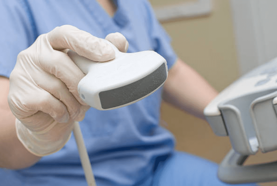
What is an ultrasound scanner, and why is it so important in modern medicine?
Ultrasound scanners provide non-invasive, real-time imaging using high-frequency sound waves to visualize internal organs, tissues, and blood flow. This technology allows healthcare professionals to diagnose, monitor, and even treat various medical conditions without resorting to surgery.
While most people are familiar with ultrasound in pregnancy, there’s a wide array of uses that go beyond this common application. Let’s explore how ultrasound scanners are applied in different fields, improving patient outcomes and saving lives.
1. Prenatal Care: More Than Just Gender Reveal
One of the most widely recognized uses of ultrasound scanners is in prenatal care. It allows us to monitor the health and development of a fetus throughout pregnancy. This includes detecting fetal abnormalities, assessing growth, and monitoring the health of both the mother and the baby.
Why is ultrasound preferred in prenatal care?
Ultrasound machine provide a safe, non-invasive way to get a real-time look at the baby’s development. This technology has reduced the need for more invasive procedures, ensuring the well-being of both the mother and the child throughout the pregnancy journey.
2. Cardiology: Seeing the Heart Like Never Before
Ultrasound scanners are also widely used in cardiology. Echocardiography, a specialized form of ultrasound, allows doctors to visualize the heart’s structure and function. This tool is crucial for diagnosing conditions like heart disease, valve dysfunction, and congenital heart defects.
What makes ultrasound ideal for heart imaging?
The ability to capture real-time images of the heart’s movement and blood flow makes ultrasound machine an essential tool in cardiology. Unlike X-rays or CT scans, they do not expose patients to ionizing radiation, making them safer for routine monitoring of heart conditions.
3. Emergency Medicine: Speed Saves Lives
In emergency rooms, speed is everything, and ultrasound scanners are vital in trauma care and rapid diagnosis. Whether it’s assessing internal bleeding, collapsed lungs, or other critical injuries, ultrasound can provide instant insights that guide life-saving decisions.
How is ultrasound used in trauma cases?
The FAST exam (Focused Assessment with Sonography for Trauma) is one of the key ultrasound techniques used to quickly identify internal injuries in trauma patients. This can be the difference between life and death when seconds matter.
4. Musculoskeletal Imaging: Not Just for Broken Bones
When people think of imaging bones, they often think of X-rays. However, ultrasound machine are increasingly used in musculoskeletal imaging, especially for soft tissues like muscles, ligaments, and tendons.
Why use ultrasound for musculoskeletal injuries?
Ultrasound offers a dynamic, real-time view, allowing doctors to assess soft tissue injuries while the patient moves, which is something static imaging techniques like MRI and X-rays cannot provide. It’s particularly valuable for athletes and people recovering from surgery.

5. Abdominal Imaging: Beyond the Surface
Ultrasound scanners are instrumental in examining the abdomen to assess organs like the liver, kidneys, pancreas, and gallbladder. They are often used to detect gallstones, liver disease, or kidney abnormalities. It’s a critical diagnostic tool for both routine checkups and urgent medical cases.
How do ultrasound scanners help in abdominal imaging?
Abdominal ultrasound is a quick, non-invasive way to diagnose internal conditions, which can prevent unnecessary surgeries or guide doctors toward the right treatment. Its use in detecting abnormalities in real-time is indispensable for healthcare providers.
Frequently Asked Questions About Ultrasound Scanners
- How do ultrasound scanners work without radiation?
- Why are ultrasound machine considered safer for pregnant women?
- Can ultrasound machines detect cancer?
- What’s the difference between an ultrasound and a CT scan?
- How accurate is ultrasound in diagnosing heart conditions?
- Is there a risk of overuse in ultrasound imaging?
These questions often come up when discussing the versatility of ultrasound technology. It’s always important to consult with a healthcare provider to understand the best diagnostic approach for individual cases.
Conclusion: A Tool for Every Field
The application of ultrasound scanners goes beyond what many expect. From prenatal care to emergency rooms, musculoskeletal imaging, and abdominal scans, ultrasound technology has reshaped the way we practice medicine. Whether you’re in cardiology or sports medicine, ultrasound scanners are a must-have in any modern healthcare setting.
By understanding the full capabilities of this technology, healthcare professionals can deliver more precise, non-invasive care, ultimately improving patient outcomes.
Stay ahead of the latest advancements and explore the potential of ultrasound scanners for your medical practice!


