

What Are the Key Differences Between 2D, 3D, and 4D Ultrasound Imaging?
Choosing between 2D, 3D, and 4D ultrasound imaging can be daunting. Each technology offers unique advantages for medical diagnostics, from prenatal care to cardiovascular assessments. By understanding their differences, industries and individuals can make informed decisions that fit their diagnostic needs.
2D imaging is a reliable diagnostic tool, 3D adds depth for structural clarity, and 4D delivers real-time motion imaging. These advancements are transforming medical diagnostics across industries, making them indispensable tools.
While 2D, 3D, and 4D imaging have distinct features, their applications often overlap in medical and non-medical fields. To choose the right technology, it’s essential to explore how each method works and what makes it suitable for different applications.
Why Is Ultrasound Imaging the Gold Standard in Prenatal Care?
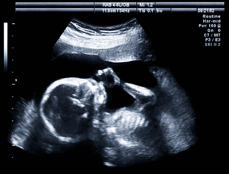
Expecting mothers rely on ultrasound imaging for reassurance and accurate monitoring. But why is 2D ultrasound still considered the gold standard for prenatal diagnostics?
2D imaging provides real-time, clear, and safe images of a fetus’s development, ensuring effective monitoring throughout pregnancy.
2D imaging is widely used in prenatal care because of its proven reliability and safety. It enables healthcare providers to monitor fetal growth, assess amniotic fluid levels, and detect any abnormalities in real-time. This imaging method is invaluable for observing the fetus’s heartbeat, position, and overall health. Additionally, 2D ultrasound is cost-effective and widely available, making it the preferred choice for routine pregnancy monitoring.
What Makes 3D Ultrasound Imaging Safe and Effective for Structural Diagnosis?
For detailed anatomical visualization, 3D ultrasound imaging offers unparalleled clarity. But how does it ensure accuracy while maintaining patient safety?
3D imaging combines multiple 2D images to create lifelike, volumetric visuals, enhancing diagnostic precision without compromising patient safety.
3D imaging provides three-dimensional images by compiling 2D slices into a volumetric dataset. This technology is particularly valuable in identifying structural abnormalities, such as fetal anomalies, organ malformations, and musculoskeletal conditions. Its enhanced visualization capabilities make it an excellent tool for preoperative planning and detailed anatomical assessments. Like 2D ultrasound, it employs non-ionizing sound waves, ensuring that it remains safe and effective for patients.
How Does 4D Ultrasound Imaging Improve Real-Time Diagnostics?

Imagine watching a moving fetus or a functioning heart in real time. That’s the power of 4D ultrasound imaging.
4D ultrasound imaging captures motion in real-time by adding a time dimension to 3D images, enhancing the ability to diagnose dynamic conditions.
4D ultrasound is an extension of 3D imaging, introducing the time factor to create real-time videos of internal movements. This technology is especially beneficial in obstetrics, where it allows healthcare professionals to observe fetal movements, facial expressions, and behaviors. Similarly, in cardiology, 4D imaging is used to visualize and assess heart function dynamically, aiding in the diagnosis and treatment of complex cardiovascular conditions. The ability to provide real-time feedback also makes it ideal for guiding interventional procedures with precision.
Which Ultrasound Imaging Techniques Are Right for Your Needs?
Your choice of ultrasound imaging depends on factors like diagnostic requirements, applications, and budget. Which option is best for you?
2D is cost-effective for routine diagnostics, 3D excels in anatomical clarity, and 4D is ideal for dynamic studies. Understanding your needs is key to making the right choice.
Selecting the right imaging technique depends on your specific requirements. For routine diagnostic needs, 2D ultrasound remains the most affordable and accessible option. For detailed anatomical assessments, 3D ultrasound offers unparalleled clarity. If dynamic, real-time visualization is required, such as for fetal movements or cardiac function, 4D ultrasound is the best choice. Understanding the unique capabilities of each method ensures precise and effective diagnostics tailored to your needs.
Where Can Ultrasound Imaging Be Used Beyond Healthcare?
Did you know that ultrasound imaging has uses far beyond hospitals and clinics? Its versatility spans multiple industries.
Ultrasound imaging is used in veterinary medicine, industrial testing, and art restoration, showcasing its versatility beyond traditional healthcare applications.
While imaging is predominantly associated with medical diagnostics, its applications extend to other fields. In veterinary medicine, it is commonly used for diagnosing conditions in animals. In industrial settings, imaging plays a critical role in non-destructive testing, identifying flaws in materials like metal and concrete without causing damage. Additionally, ultrasound technology is used in art restoration, allowing experts to analyze and preserve historical artifacts and paintings without altering them. These diverse applications highlight the adaptability and innovation of ultrasound technology.
2D, 3D, and 4D ultrasound imaging each bring unique strengths to the world of diagnostics. Whether it’s affordability, anatomical precision, or dynamic real-time imaging, these technologies are revolutionizing diagnostics across industries. By understanding their differences, individuals and organizations can confidently choose the right tool to meet their goals.


